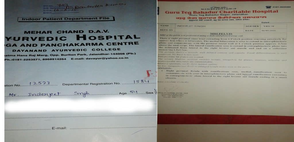
Case Study: Inderjeet Singh

Patient Name: Inderjit Singh
Age: 54/M
MRI Pelvis
There is right perineal sinus tract extending from 6 o’clock position coursing anteriorly for approximately 4 cm where it bifurcates. The medial bifurcation is seen to extend in intersphincteric plane and communicate with the posterior renal wall at 6 o’clock position approximately 2.5 cm above the anal verge. The lateral ramification seen to extend in extra sphincteric plane into the issioanal fossa lateral to the right levator ani muscle and end on a collection measuring 1.5 x 1.5 cm.
The prostate shows a smooth well-defined contour and capsule zonal differentiation is well preserved. No focal lesions seen periprosthetic fat planes are normal. The unary bladder, seminal vesicles, rectum, intra pelvic fat planes, osseous articular and neuro vascular structures are unremarkable. Bilateral spermatic cord show normal outline. No free interperineal fluid or enlarged lymph nodes seen in the pelvis
Opinion:
Fistula with ramification seen. Medial ramification seen to communicate with anus in inter sphincter plain and lateral ramification extending an extra sphincteric plane lateral to the right levator and ani muscle ending in a small abscess.